
QUT Analysis in Focus: Take a look at 2022 finalists

[ad_1]
The finalists of QUT’s annual Analysis in Focus multimedia competitors have as soon as once more lit the college’s analysis variety in artistic mild.
The winners of the 2022 competitors can be introduced this Thursday at a showcase at The Dice (4:30pm-6pm) on the Gardens Level campus, together with the Individuals’s Alternative award (common). choose by way of Fb ends at 11.59 p.m. Tuesday).
A panel of judges chosen this 12 months’s entries to pick the next 10 finalists.


2022 QUT Examine in Finalist Highlight
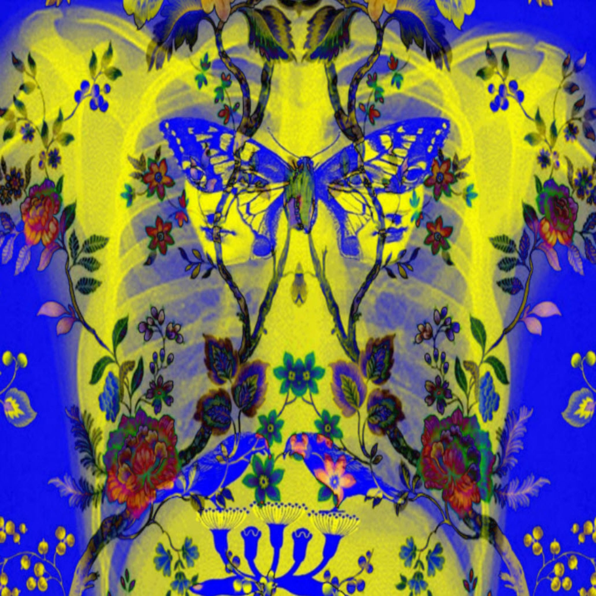
Dusan Bojic – SpiroArtis Picture: This art work was created from the breath of an adolescent affected person with a respiratory sickness, utilizing SpiroArtis – the primary art-based interactive well being expertise platform on the planet. world, was based by QUT Doctorate in Inventive Industries pupil Dusan Bojic as an progressive approach so as to add momentum to lung quantity testing.
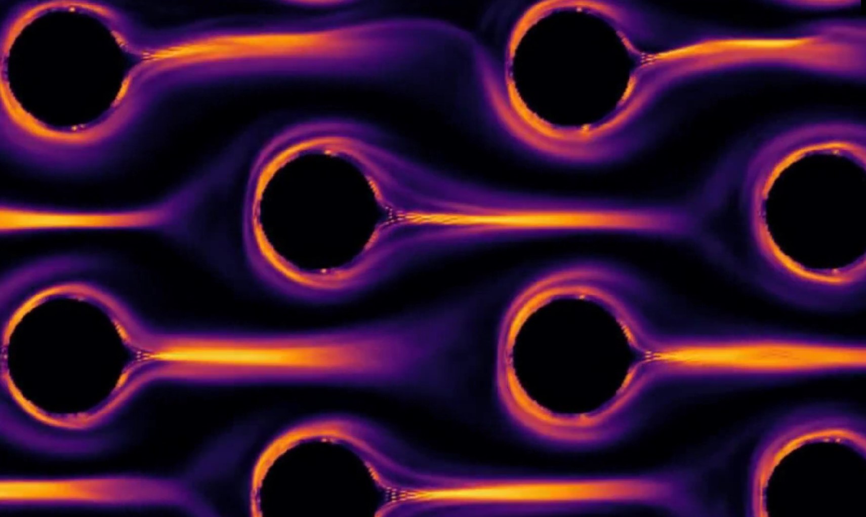
Vedad Dzanic – The Hypnotic Dance of Polymers: Animation displaying numerical simulation of viscous fluid (a solvent liquid blended with polymer components) flowing by a cylindrical impediment. When the polymer is overstretched, the viscoelastic fluid turns to a chaotic state, and an elastic chaos is fashioned. (Click on the picture beneath to observe the video.)
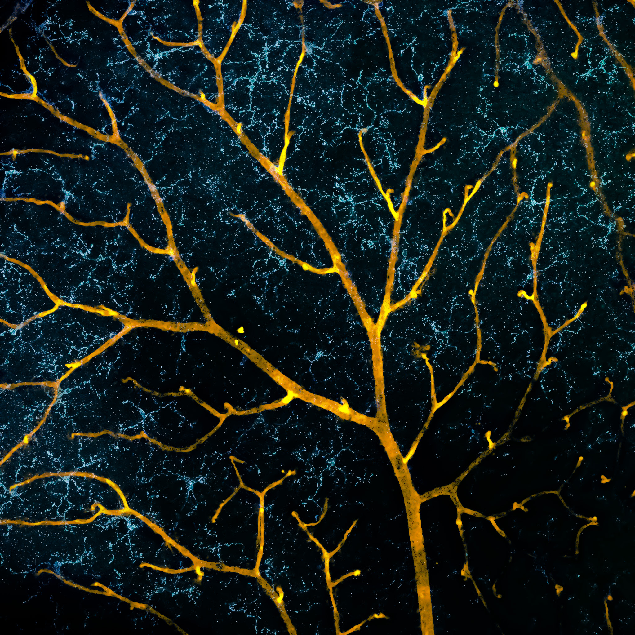
Fazeleh Etebar – Dizziness: It is a confocal fluorescence microscopy picture of a mouse retina displaying a layer in the back of the attention. The colours signify the vascular system (yellow) and microglia (blue).
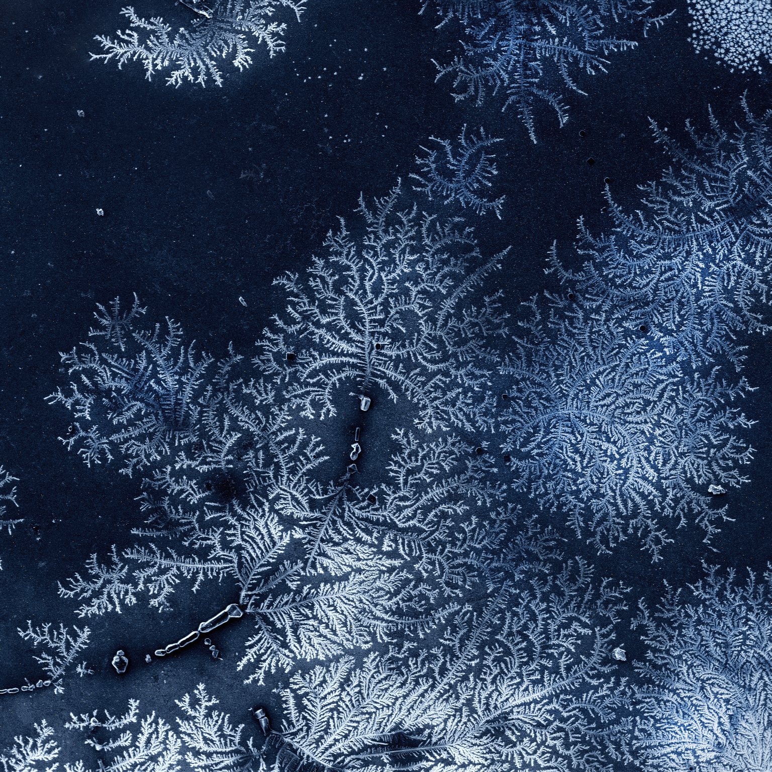
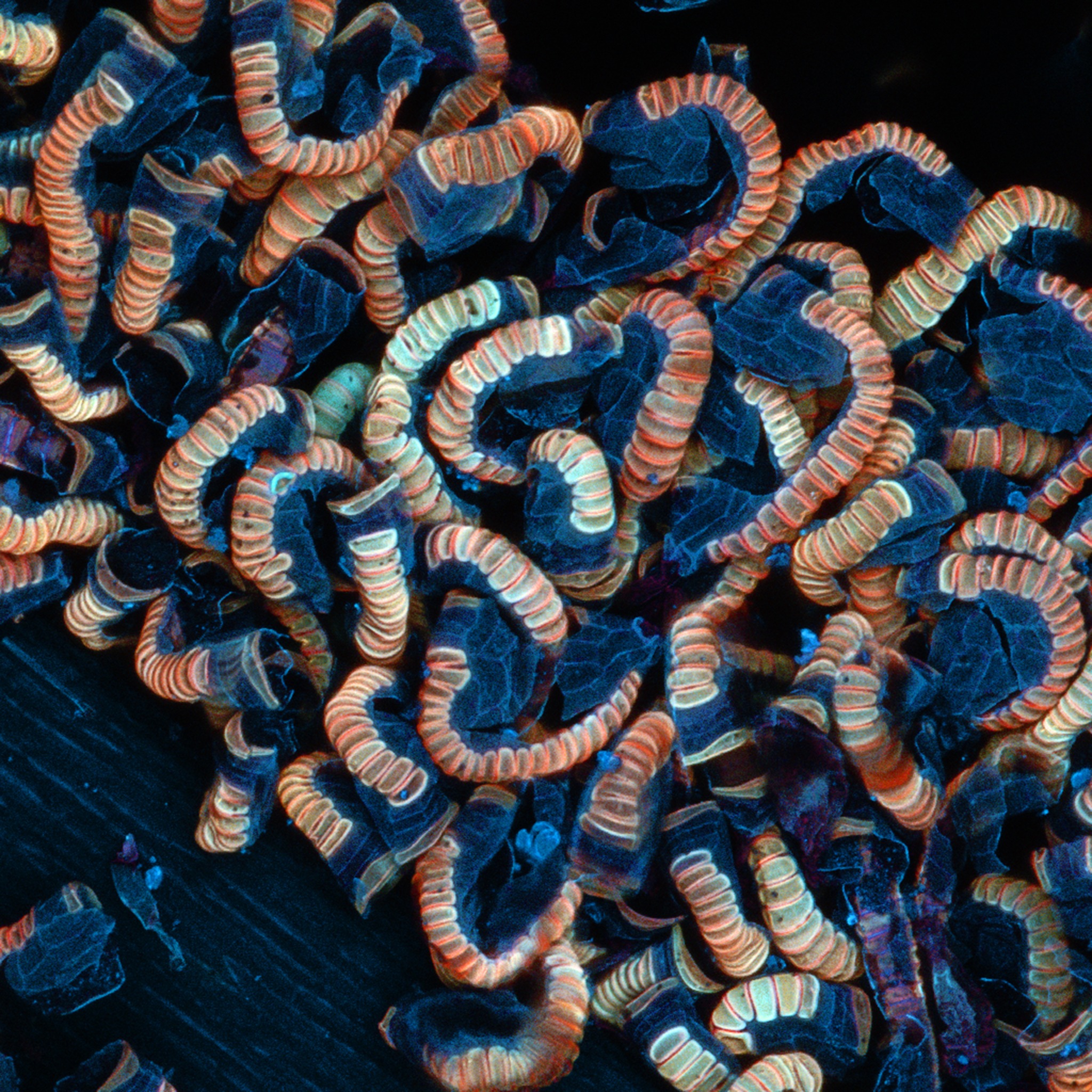
Konstantin Faershteyn – Ambient solidified fractals: The speedy solidification of salts typically produces the attribute tree-like construction of the crystal – the dendritic construction – as this scanning electron microscope picture depicts. This polymorphic kind can also be typical for snowflakes or the so-called “frozen fractal”.
John Griffin – Subsequent Era: That is a picture of a fern at QUT’s Gardens Level campus, displaying the pure fluorescence of the pattern underneath the microscope’s laser. The picture demonstrates the capabilities of one in all QUT’s superior imaging platforms.
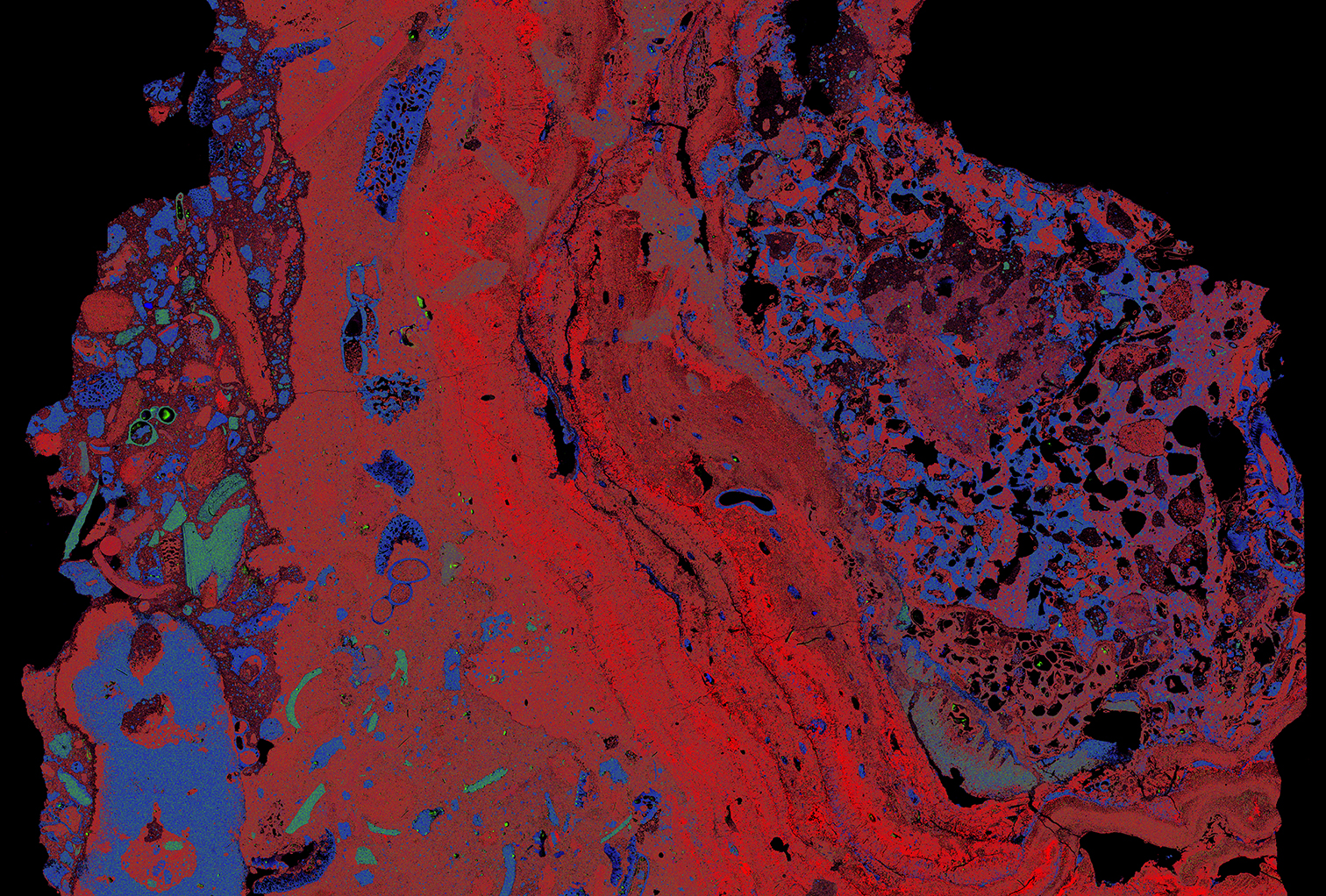
Lisa Kearney – Preserve It Collectively: It is a multicomponent map of reef rock produced with a Tescan TIMA scanning electron microscope at QUT’s Central Analytical Analysis Facility. TIMA generates massive space maps that can be utilized to look at patterns at totally different scales. Like analysis college students, coral reefs typically attempt to maintain it collectively, discovering assist within the distinctive group that surrounds them.
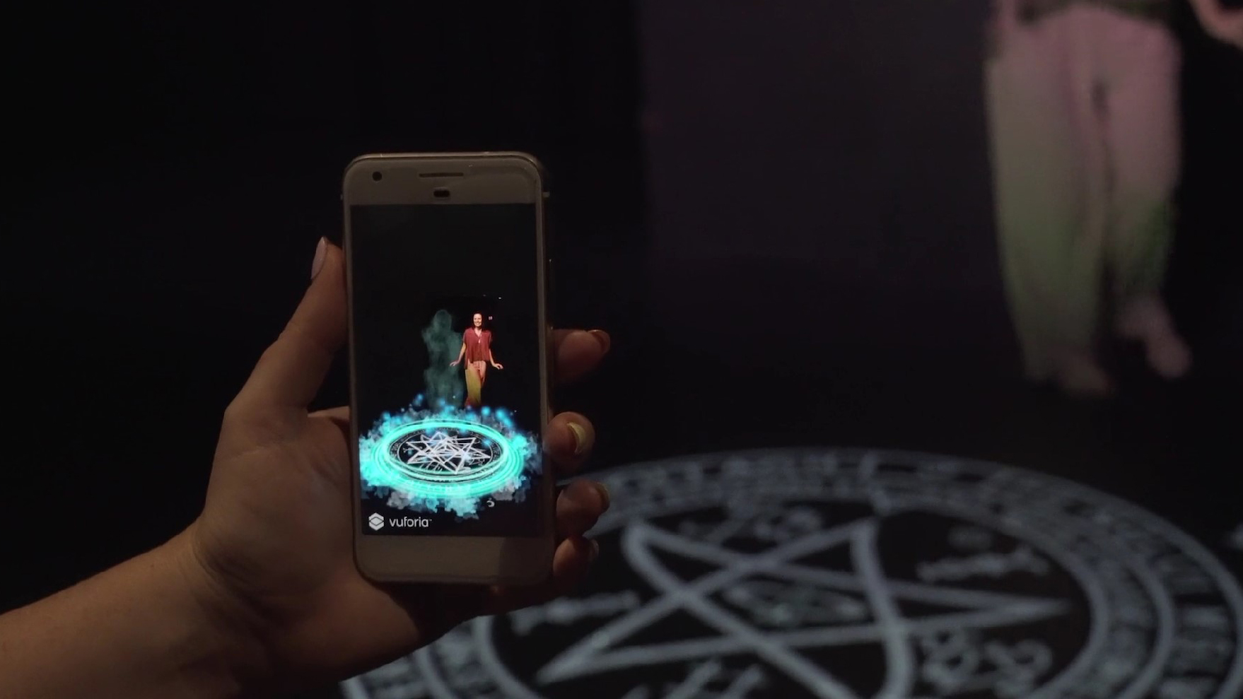
Kristen Maloney – Ghost Story: It is a cross-industry collaboration between efficiency and laptop programming to create a efficiency that enhances creativity. This new type of digital efficiency integrates stay efficiency and augmented actuality and is skilled by a smartphone app. (Click on the picture beneath to observe the video.)
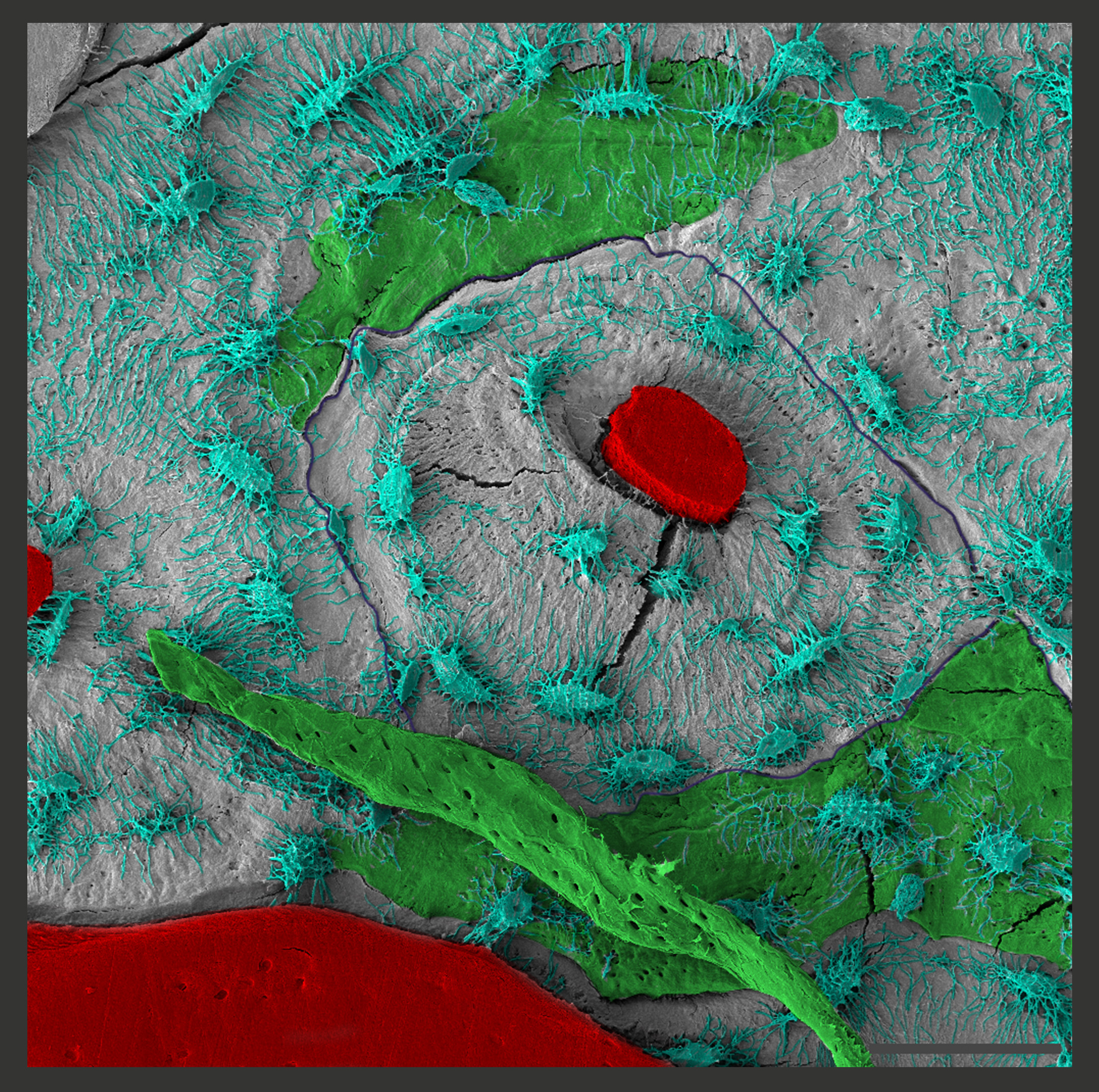
Flavia Medeiros Savi – Bone Focal: This scanning electron microscope picture exhibits the ‘area of curiosity’ of a 3cm tibia defect, reconstructed with a medical-grade polycaprolactone holder mixed with a corticoperiosteal flap. The picture exhibits new bone tissue being constructed and the community of bone cells distributed across the spinal canal.
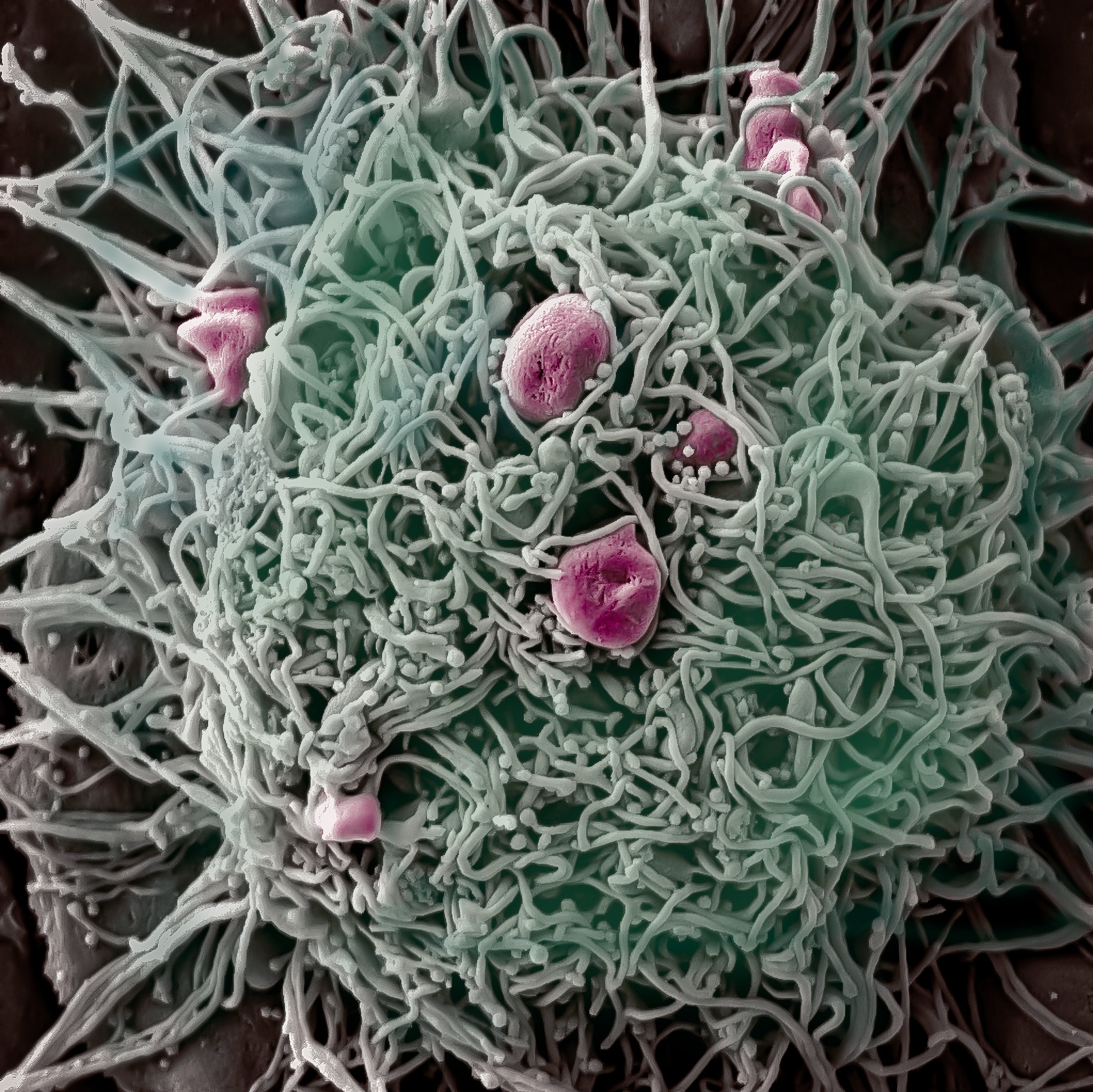
Jayanti Mendhi – Go hungry however do not be silly: Macrophages are a kind of immune cell often known as ‘phagocytosis, a phrase of Greek origin which means ‘cell that eats or swallows’. That is an electron microscope picture displaying micro organism (in pink) engulfed by an activated macrophage cell. The goal of this examine was to learn the way an antimicrobial coating that efficiently kills micro organism impacts the power of macrophages to eat these micro organism.
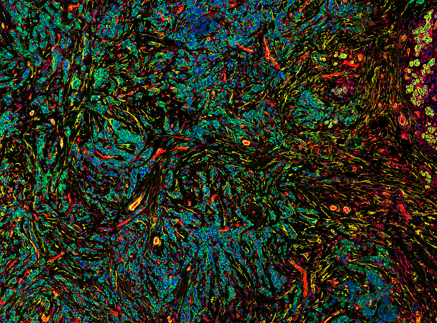
Akhilandeshwari Ravichandran – Illuminating Most cancers: This GeoMx digital spatial profile scan exhibits the tumor area of the tissue part of a triple destructive breast most cancers affected person, illuminated utilizing morphological markers fluorescence – PanCK (inexperienced, most cancers cells), DNA (blue, all cells), SMA (yellow) and CD31 (purple, endothelial cells).
[ad_2]