
Designing an Ultrasound Sticker that may Picture your Inside Organs
[ad_1]
On this interview, we discuss to researchers from the Zhao lab at MIT about their new ultrasound sticker that may present non-invasive imaging of inside organs for as much as 48 hours.
Please introduce your self and inform us what impressed your newest analysis?
We’re a group of engineers from MIT Zhao lab (http://zhao.mit.edu/)
Present medical analysis primarily depends on medical radiology instruments for imaging organs to make selections. Nevertheless, these are sometimes rare medical approaches and can most certainly miss illness types. To deal with this problem, we goal to develop a wearable that may present long-term imaging capabilities for each clinicians and sufferers, monitoring their sickness or well being situation.


In healthcare settings, clinicians usually want photos of a affected person’s inside organs. To do that, ultrasound imaging is usually used. Are you able to inform us extra about how ultrasound imaging works?
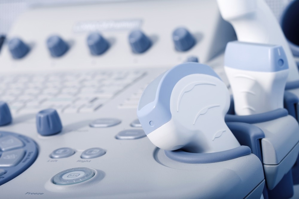
Ultrasound imaging is a secure and non-invasive window into the workings of the physique, offering clinicians with direct photos of a affected person’s inside organs. To seize these photos, skilled technicians manipulate ultrasound wands and transducers to direct sound waves into the physique. These waves are mirrored again to create high-resolution photos of the affected person’s coronary heart, lungs, and different deep organs.

Picture credit score: HOMONSTOCK/Shutterstock.com
Though ultrasound imaging has gained reputation in healthcare settings, it has many disadvantages. What are a few of these drawbacks, and the way does your new know-how assist overcome them?
Often, for ultrasound imaging, the technician first applies a liquid gel to the affected person’s pores and skin, which transmits ultrasound waves. A transducer, or transducer, is then pressed into the gel, sending sound waves into the physique that bounce off inside constructions and again to the transducer, the place the echoes are transformed into visible photos.
For sufferers requiring long-term imaging, some hospitals supply probes hooked up to robotic arms that may maintain the transducer in place with out fatigue, however the liquid ultrasound gel will soften and dry. over time, disrupting the long-term imaging course of.
To unravel this downside, we designed an ultrasound patch that produces larger decision photos for an extended time by coupling the stretchy adhesive to a inflexible array of transducers. This mix permits the gadget to evolve to the pores and skin whereas sustaining the relative place of the transducers to supply clearer and extra correct photos.
In your newest analysis, you designed a brand new ultrasound sticker. How did you design this sticker and the way does it work?
We have designed a brand new ultrasound patch that produces high-resolution photos for an extended time frame by pairing a stretchy adhesive with a inflexible array of transducers. This mix permits the gadget to evolve to the pores and skin whereas sustaining the relative place of the transducers to supply clearer and extra correct photos.
The gadget’s adhesive layer is manufactured from two skinny layers of elastomer encased in a strong hydrogel within the center, a principally water-based materials that readily transmits sound waves. The MIT group’s hydrogel is elastic and stretchy, not like conventional ultrasound gels. The elastomer prevents water lack of the hydrogel. When the hydrogel is extremely hydrated, sound waves can penetrate effectively and provides excessive decision photos of inside organs.
The underside elastic layer is designed to stay to the pores and skin, whereas the highest layer sticks to a inflexible array of probes that the group additionally designed and constructed. All the ultrasonic sticker is about 2 sq. centimeters throughout and three mm thick – the dimensions of a postage stamp.
To check our design, we handed the ultrasonic patch by means of a check battery with wholesome volunteers, who wore the patch on completely different elements of the physique, together with the neck, chest , stomach and arms. The patches remained hooked up to their pores and skin and produced clear photos of the underlying constructions for as much as 48 hours.
Ultrasound stickers
Your ultrasound patch may present steady photos of inside organs for 48 hours, in addition to document photos because the affected person performs numerous actions. How is that this attainable, and what benefit does it have for medical doctors by observing organs over a time frame versus a single snapshot second?
Our BAUS system permits this and an adhesive ultrasound patch may be utilized to human pores and skin, conserving stability and visualization of inside organs steady. An imaging gadget that maintains steady monitoring of particular physique elements can be utilized to observe and diagnose numerous illnesses. Medical doctors can intently monitor tumor development over time.
Somebody at excessive danger for hypertension can put on an ultrasound patch to measure their hypertension, alert them to a spike in stress, or monitor if a drugs is working. A COVID affected person can keep at residence, realizing that an imaging gadget will alert them if their sickness causes a lung an infection so extreme that it requires hospitalization.
Do you hope that with continued analysis on these ultrasound patches, they are often made accessible in pharmacies in order that sufferers needn’t go to a healthcare facility? How does this profit each the affected person and the physician?
Sure, we hope the BAUS may be one of many future health trackers that may be purchased on the pharmacy. Due to this fact, we’re designing mass manufacturing capabilities to chop prices even additional, with the worth per sticker at present round $200. The large promoting/benefit of this new gadget is that it opens up new kinds of medical diagnoses that aren’t attainable in a static surroundings.
For instance, to evaluate coronary heart well being, measuring organ exercise throughout train could be useful – however it’s troublesome to carry the ultrasound wand in opposition to the goo-covered chest of a operating topic. With wearable ultrasound patches, the place you do not have to maintain the transducer in your physique, they may present you could get high-quality photos of your coronary heart even whereas in movement. This know-how can ease the burden on each medical doctors and sufferers.

Picture credit score: Felice Frankel/Shutterstock.com
Medical wearables have acquired rising consideration lately, thanks partially to synthetic intelligence and machine studying. How can each of those guidelines be utilized to your ultrasonic stickers for additional improvement?
An enormous path that we’re taking is the appliance of synthetic intelligence to the BAUS system. A picture is simply priceless in case you can truly diagnose it. So even when we are able to get all these photos, we nonetheless need assistance to get helpful medical diagnoses from them.
We’re constructing algorithms that may monitor organ physiology, quantitatively analyze it, and arrive at diagnostic selections. This may scale back the burden on the clinician.
Do you consider your know-how might help enhance medical diagnoses worldwide? What might this imply for world well being?
A very powerful software we envision might be the detection and analysis of coronary heart assaults. Coronary heart well being is on the radar of different wearable builders. For instance, smartwatches just like the Apple Watch have the power to observe electrical indicators that point out coronary heart exercise utilizing a so-called electrocardiogram (ECG or EKG). This can be utilized to diagnose coronary heart assaults—not less than in some circumstances.
Research present that an EKG can solely diagnose about 20% of coronary heart assaults. Nearly all of true coronary heart assaults require imaging modalities, equivalent to ultrasound imaging, for analysis. Steady imaging of a affected person’s coronary heart can seize their signs and supply an early analysis. We envision that ultrasound patches might be packaged and bought by sufferers and shoppers and used not solely to observe numerous inside organs but additionally tumor development. , in addition to fetal improvement within the womb.
Your analysis is funded partially by MIT but additionally by numerous organizations, together with the NIH, the US Military Analysis Workplace, and the Nationwide Science Basis. How vital is funding in discovering new scientific applied sciences?
Funding is crucial to allow understanding of fundamental science and the event of know-how that may deal with main societal challenges. We drastically recognize the varied funding companies which have trusted and supported our work.
What are the subsequent steps for you and your analysis?
We’re engaged on a completely built-in BAUS system that anybody can use, and we’re additionally working with medical doctors to gather medical information and put the gadget into medical trials. .
The place can readers discover extra info?
Our group’s web site: http://zhao.mit.edu/
About Professor Xuanhe Zhao
Xuanhe Zhao is a professor of mechanical engineering and civil and environmental engineering (courtesy) at MIT. Zhao Lab’s mission at MIT is to advance the science and know-how of the human-machine interface to deal with the foremost societal challenges of well being and sustainability with built-in experience in mechanics, supplies supplies and biotechnology. The primary focus of Zhao Lab’s present analysis is the analysis and improvement of sentimental supplies and programs. 
About Chonghe Wang
Chonghe Wang is at present a graduate pupil working with Professor Xuanhe Zhao on the Division of Mechanical Engineering of the Massachusetts Institute of Expertise. Chonghe has greater than 10 years of analysis expertise in ultrasonic know-how.
Since 2016, he has pioneered the know-how of wearable ultrasound know-how that may monitor very important indicators of deep tissue within the human physique. His main analysis has been printed in Science (2022), Nature Biomedical engineering (2021), Nature Biomedical engineering (2018) and has been solely featured by lots of of well-known media, together with: Nationwide Geographic Journal, Forbes, MIT Expertise Overview, Nationwide Institute of Well being (NIH), and lots of others. This know-how has big potential in remodeling the paradigm of changing cumbersome medical ultrasound machines into wearable sensible units for next-generation digital healthcare.
His works have been accepted and printed by a collection of well-known journals equivalent to Nature, Science, Nature Biomedical Engineering, Nature Nanotechnology, Nature Electronics, Science Advances, Science and Expertise. Proceedings of the Nationwide Academy of Sciences, Superior Supplies, and lots of different journals. He was acknowledged because the ‘2019 Baxter Younger Investigator’ for his contributions in growing the world’s first wearable central sphygmomanometer able to saving ICU sufferers’ lives.
About Dr. Xiaoyu Chen
Xiaoyu Chen is a postdoctoral fellow within the Division of Mechanical Engineering on the Massachusetts Institute of Expertise. He acquired his Bachelor’s diploma from the School of Chemistry at Jilin College in 2013. He obtained his Ph. graduated from the Division of Biomedical Engineering on the Chinese language College of Hong Kong in 2019. His analysis focuses on the design and preparation of polymeric biomaterials for biomedical functions. He has acquired the CUHK Distinguished Scholar Award and the Biomaterials Affiliation STAR Award.
[ad_2]


